Overview
Electrical impedance tomography (EIT) has been studied to monitor lung ventilation because it is the only real-time lung imaging method without large equipment. However, previous EIT systems just provided 2D cross-sectional image with limited spatial information of the lung and unneglectable volume detection error depending on the location of 2D EIT belt relative to the patient’s lung. In spite of its importance, the 3D-EIT has not been realized in lung monitoring because it has many design challenges such as noises incurred by complicated wiring, long cable length, wide variation in electrode contact and signal, and large personal-to-person impedance variation. In this paper, we present a portable 3D-EIT SoC for real-time lung ventilation monitoring with following 5 features: 1) The active electrodes (AEs) system to reduce coupling noise, 2) High output impedance current stimulator to inject stable current, 3) Impedance spectroscopy to enable both time-difference (TD) EIT and frequencydifference (FD) EIT, and to select an optimal frequency for TD-EIT, 4) Wide-dynamic range front-end circuit to detect variable ranges of signal with highinput impedance and CMRR, 5) Calibration to reduce the electrical characteristics variations of AEs.
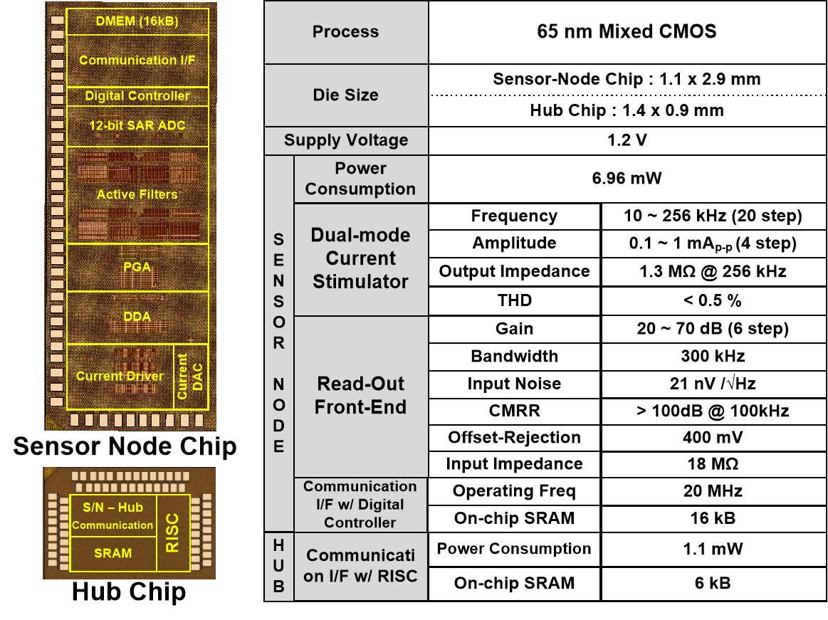
Implementation results
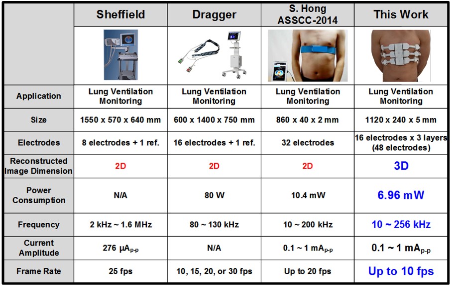
Performance comparison
Architecture
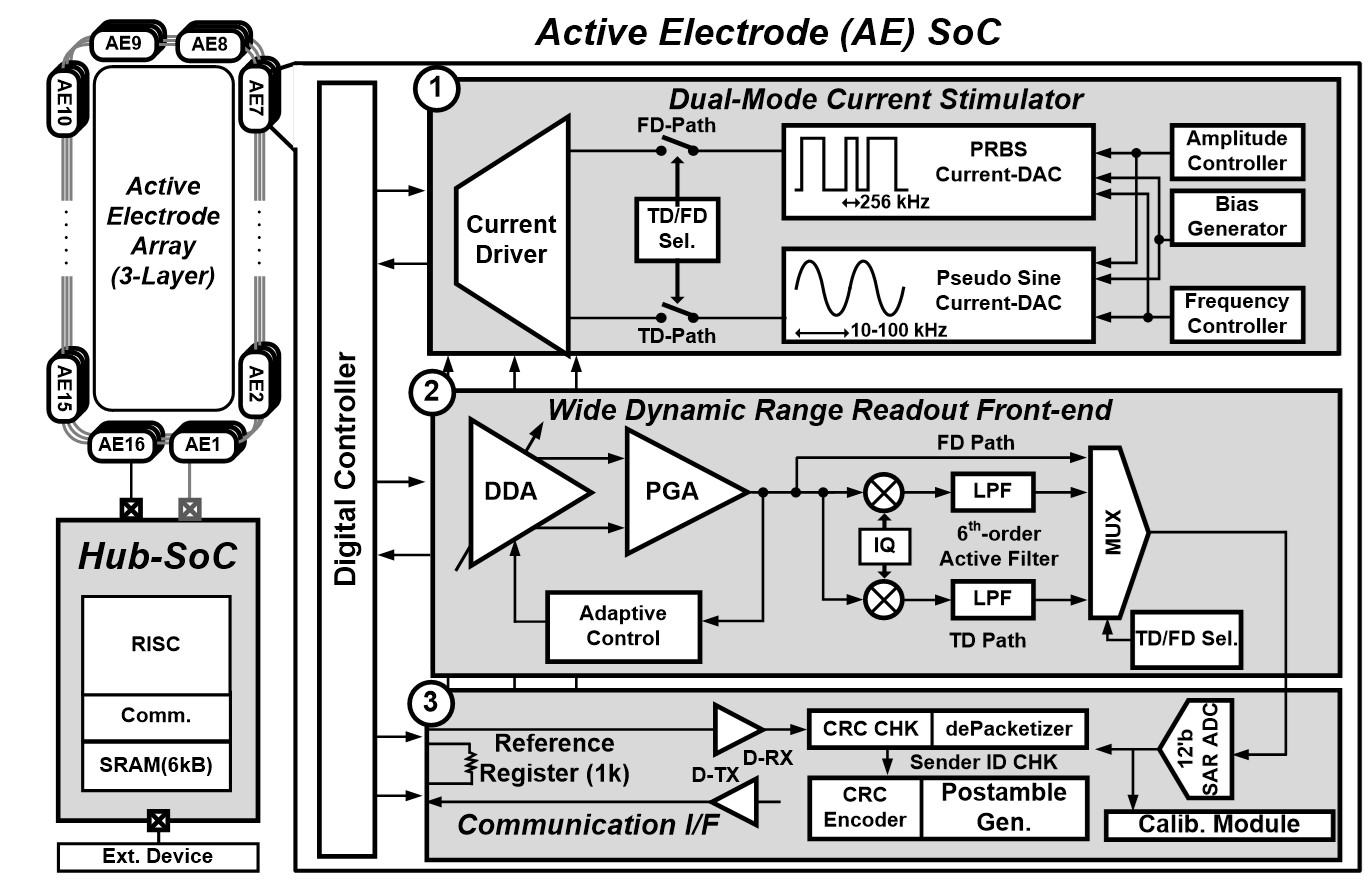
Features
- Dual-mode Current Stimulator
- Wide Dynamic Range Readout Front-end
- Calibration Module with Auto Calibration ADC
Related Papers
- ISSCC 2017 [pdf]